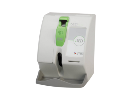
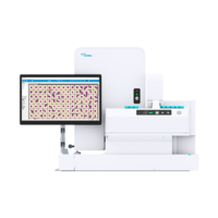
DI-60
![[.CH-fr Switzerland (french)]](/fileadmin/_processed_/6/6/csm_DI-60-3_c562052b61.png "[.CH-fr Switzerland (french)]")
![[.CH-fr Switzerland (french)]](/fileadmin/_processed_/4/9/csm_DI-60-1_fe9ee33edf.png "[.CH-fr Switzerland (french)]")
![[.CH-fr Switzerland (french)]](/fileadmin/_processed_/6/6/csm_DI-60-3_4cf0051ecd.png "[.CH-fr Switzerland (french)]")

![[.CH-fr Switzerland (french)]](/fileadmin/_processed_/4/9/csm_DI-60-1_7c7e4c73dc.png "[.CH-fr Switzerland (french)]")
Sysmex DI-60 - Rapide et précis, fiable et en réseau
- Révision et validation efficaces et détaillées pour une plus grande précision
- Des flux de travail plus performants et plus rapides
- Des temps de traitement plus courts grâce à la détection automatique
- Stockage et archivage à long terme des images cellulaires
- Cohérence dans la qualité de l'analyse
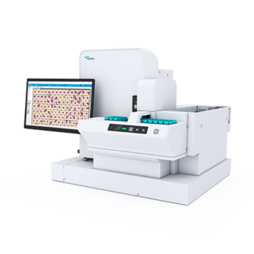
L'analyseur morphologique numérique Sysmex DI-60 rime avec efficacité, qualité et sérénité. Il s'agit du seul dispositif d'analyse numérique sur le marché, entièrement automatisé et intégré au flux de travail en hématologie, directement raccordé au réseau. En d'autres termes, les temps de traitement sont plus rapides qu'avec les appareils traditionnels, car vous n'avez plus besoin de transporter manuellement les lames vers l'analyseur. Vous disposez de temps libre supplémentaire pour réaliser des tâches plus pertinentes. Les problèmes de standardisation, de qualité et de risque biologique appartiennent au passé. Supporté par le service et l'assistance réputés de Sysmex, cet appareil fournit un excellent niveau de qualité d'analyse et de détails auquel vous pouvez vous fier. Rapidité, qualité, sécurité et efficacité en un seul analyseur.
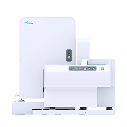
Le Sysmex DI-60 est un système d'analyse d'imagerie cellulaire automatisé. Il est directement raccordé au système de détection de l'analyseur et élimine par conséquent le besoin d'une intervention manuelle dans le flux de travail en hématologie durant le cycle d'imagerie. Le dispositif lui-même se compose d'un microscope automatique, d'un appareil photo numérique d'une très grande qualité et d'un système informatique qui collecte et procède à un classement préalable des cellules obtenues à partir des frottis sanguins colorés. Il localise automatiquement les cellules sur la lame et prend des photographies de chaque cellule trouvée ; après quoi il les analyse et effectue un pré-classement au moyen d'un traitement d'images avancé. Le nombre de leucocytes analysés peut être défini par l'utilisateur.
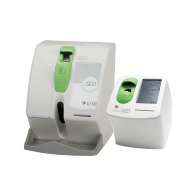
This is what the DI-60 consists of
The Sysmex DI-60 is an automated, cell-locating image analysis system. It is connected directly to the analyser track and therefore eliminates the need for manual intervention in the haematology workflow in the imaging cycle.
The device itself consists of an automated microscope, an extremely high quality digital camera and a computer system that collects and pre-classifies cells from stained blood smears. It automatically locates cells on the slide and takes an image of each cell found, after which it analyses and pre-classifies them using advanced image processing. The number of white blood cells that are analysed is user-definable.
This is how the DI-60 works
To perform a differential count with morphology assessment, a thin film of blood is wedged on a glass slide from a peripheral blood sample and stained according to the RAL MCDh, May-Grünwald Giemsa or Wright protocol. With XN, this is done fully automatically by the SP-10 slide maker and stainer module. The DI-60 analyser acquires and pre-classifies the cells, after which the operator verifies and modifies the suggested classification of each cell if necessary. The operator may also introduce additional observations and comments when needed.
This is what the DI-60 analyses
The DI-60 can pre-classify 12 WBC categories. These include segmented and band neutrophils, eosinophils, basophils, monocytes, lymphocytes, atypical lymphocytes, plasma cells, promyelocytes, myelocytes, metamyelocytes, and blast cells. In addition to white blood cells, the system also pre-classifies the following five non-WBC categories: smudge, artefacts, giant platelets, platelet clumps and nucleated red cells. These are reported as the number of NRBC/100 WBC. Cells that are pre-classified with a low confidence level are placed in a category called “Unidentified.”
The device also presents an overview that can be used to characterise red blood cell morphology. Thirty-five images are collected in the optimal RBC examination area for pre-characterising the following characteristics: polychromasia, hypochromasia, anisocytosis, macrocytosis, microcytosis, and poikilocytosis. The system also provides tools that let the user improve the efficiency and consistency of estimating the PLT concentration. All images and results are stored in a database.
| Technology | Artificial Neural Network |
| Slide identification | Barcode labelled slides |
| Storage capacity | Up to 4000 slides per database |
| Supported Stains | Romanowsky stains
|
| WBC Pre-classification | segmented and band neutrophils, lymphocytes, monocytes, eosinophils, basophils, metamyelocytes, myelocytes, promyelocytes, blasts, variant lymphocytes, plasma cells non-WBC: smudge, artefacts, giant platelets, platelet clumps, erythroblasts |
| RBC Pre-characterisation | aniso-, micro- and macrocytosis, poly- and hypochromasia, poikilocytosis |
| Body Fluid | segmented neutrophils, eosinophils, lymphocytes, macrophages (incl. monocytes), basophils, Other |
| Slide Scan | Scanning of user definable areas of a slide |
| Options | Sysmex Remote Review Software Sysmex Body Fluid application |
Sysmex Suisse AG
Tödistrasse 50
CH-8810 Horgen
+41 44 718 38 38
+41 44 718 38 39
Sysmex Suisse AG
Rue Galilée 15
CH-1400 Yverdon-les-Bains
+41 24 423 93 93
+41 24 423 93 98
Product documents
Safety data sheets
Regulatory Documents
Regulatory documents, such as Instructions for Use, can be accessed with a valid My Sysmex login:
Go to My Sysmex